生体フォトニクス
Biophotonics
TEL 06-6879-4945
FAX 06-6879-4946
概要
Biophotonics
The research in this lab aims to develop and extend optical technologies to widen the tool set that is currently available to immunology. The research can be divided into two main themes:
1) Label-free imaging. Raman scattering is an optical process that occurs when light hits a target (e.g. a living cell). A portion of the light energy can be absorbed by the vibrational modes of the molecules in the cell and this manifests itself as a color shift in the scattered light spectrum. By scanning the light beam through the sample and collecting and analyzing the scattered light spectra we can image the molecular distribution in the cell. Technically, the signal level is so low that this process has not been practical, but if the technical challenges are solved, this process allows completely label-free imaging of the molecular changes in the cell. In this research, we are developing methods to increase the speed, resolution and the signal-to-noise ratio of the imaging with the goal of time-resolved high-spatial resolution imaging of the immune response without using any fluorescent labels. Spectral signals of interest are typically lower than the noise level in measured spectra and we are using a combination of mathematical processing techniques and a priori information (in collaborations with other groups) to improve the imaging quality.
2) Nanoparticle assisted imaging and laser-induced modification of cell structure: Metallic nanoparticles have been show to act as local antenna for a light field, and can enhance the local field around the particle. This allows the particle to act as a probe for the local environment and since the particle size can be as small as 40 nanometers, the particle can enter a living macrophage and the combination of nanoparticle and laser light can give very high resolution Raman imaging. Raman measurements using nanoparticles have several inherent challenges, and some of these challenges are the focus of this research theme. The particle can influence the macrophage response, the laser light can heat the particle and damage the macrophage, and the location of the nanoparticle in the macrophage is typically not well controlled. These limitations are also interesting opportunities to explore new avenues, and the use of the nanoparticle heating can be used as a localized target to knock out subcellular regions and determine the cell's resistance to location-specific damage. The laser beam itself can also modify the cell, and create transient permeability changes in intracellular membranes, allowing laser generated biological reactions such as calcium waves and modification of the membrane potential. This research theme contains different sub-themes focused on laser-cell-nanoparticle interactions and applications of the new findings as tools for cell-based studies in immunology.

Contrast is generated using Raman signal wavenumber ranges mapped to color channels as follows: Red (2921 to 3023cm-1), Green (2714 to 2897cm-1), and Blue (1321 to 1646cm-1).
主任研究者
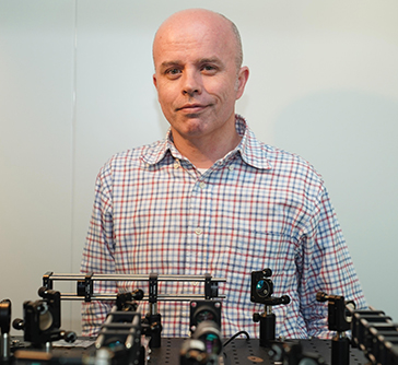
Nicholas Isaac Smith 准教授

研究内容
学歴
| 2002 | Ph.D. (Engineering/Applied Physics) Osaka University |
|---|---|
| 1997 | B.Sc., honours (Physics, 1st class Hons) University of Sydney |
| 1995 | B.Sc. (Physics/Pure Mathematics) University of Sydney |
職歴
| 2007- | Designated Lecturer, Photonics Center, Osaka University. |
|---|---|
| 2003 | Assistant Professor, Dept Frontier Biosciences, Osaka University. (-2007) |
| 2002 | JST Post-doctoral fellowship at Osaka University. (-2003) |
メンバー
- Nicholas Isaac Smith 准教授
nsmithap.eng.osaka-u.ac.jp - Nicolas Pavillon 助教(講師)
n-pavillonifrec.osaka-u.ac.jp
業績
論文
- Y. Kumamoto, K. Fujita, N. I. Smith and S. Kawata, "Deep-UV biological imaging by lanthanide ion molecular protection", Biomed. Opt. Express 7(1), pp. 158-170 (2016).
- K. Watanabe, A. F. Palonpon, N. I. Smith, L.-d. Chiu, A. Kasai, H. Hashimoto, S. Kawano and K. Fujita, "Structured line illumination Raman microscopy", Nat. Commun. 6, pp. 10095 (2015).
- N. I. Smith and N. Pavillon, "Fabrication of Gold Nanoparticles inside Cells by Laser Irradiation and their Application as Surface-Enhanced Raman Probes", レーザ加工学会誌 (Japan Laser Processing Society Journal) 22(3), pp. 228-232 (2015).
- K. Bando, N. Smith, J. Ando, K. Fujita and S. Kawata, "Analysis of dynamic SERS spectra measured with a nanoparticle during intracellular transportation in 3D", J. Opt. 17(11), pp. 114023 (2015).
- M. Yamanaka, K. Saito, N. I. Smith, Y. Arai, K. Uegaki, Y. Yonemaru, K. Mochizuki, S. Kawata, T. Nagai and K. Fujita, "Visible-wavelength two-photon excitation microscopy for fluorescent protein imaging", J. Biomed. Opt. 20(10), pp. 101202-1-101202-11 (2015).
- Y. Yonemaru, A. F. Palonpon, S. Kawano, N. I. Smith, S. Kawata and K. Fujita, "Super-Spatial- and -Spectral-Resolution in Vibrational Imaging via Saturated Coherent Anti-Stokes Raman Scattering", Phys. Rev. Appl. 4, pp. 014010 (2015).
- L.-d. Chiu, A. Palonpon, N. I. Smith, S. Kawano, M. Sodeoka and K. Fujita, "Dual-polarization Raman spectral imaging to extract overlapping molecular fingerprints of living cells", J. Biophotonics 8(7), pp. 546-554 (2015).
- N. Pavillon and N. I. Smith, "Implementation of simultaneous quantitative phase with Raman imaging", EPJ Techniques and Instrumentation 2(5), pp. 1-11 (2015).
- A. J. Hobro, N. Pavillon, K. Fujita, M. Ozkan, C. Coban and N. I. Smith, "Label-free Raman imaging of the macrophage response to the malaria pigment hemozoin", Analyst 140, pp. 2350-2359 (2015).
- N. Pavillon and N. I. Smith, "Maximizing throughput in label-free microspectroscopy with hybrid Raman imaging", J. Biomed. Opt. 20(1), pp. 016007-1-016007-10 (2015).
- N. I. Smith, K. Mochizuki, H. Niioka, S. Ichikawa, N. Pavillon, A. J. Hobro, J. Ando, K. Fujita and Y. Kumagai, "Laser-targeted photofabrication of gold nanoparticles inside cells", Nat. Commun. 5(5144), pp. 1-9 (2014).
- K.-C. Huang, K. Bando, J. Ando, N. I. Smith, K. Fujita and S. Kawata, "3D SERS (surface enhanced Raman scattering) imaging of intracellular pathways", Methods 68(2), pp. 348-353 (2014).
M. Yamanaka, N. I. Smith and K. Fujita, "Introduction to super-resolution microscopy", Microscopy (Tokyo) 63(3), pp. 177-192 (2014). - Y. Yonemaru, M. Yamanaka, N. I. Smith, S. Kawata and K. Fujita, "Saturated Excitation Microscopy with Optimized Excitation Modulation", Chem. Phys. Chem. 15(4), pp. 743-749 (2014).
- D. Pissuwan, A. J. Hobro, N. Pavillon and N. I. Smith, "Distribution of label free cationic polymer-coated gold nanorods in live macrophage cells reveals formation of groups of intracellular SERS signals of probe nanoparticles", RSC Advances 4(11), pp. 5536-5541 (2014).
- N. Pavillon, K. Fujita and N. I. Smith, "Multimodal Label-free Microscopy", J. Innov. Opt. Health Sci. 7(5), pp. 1330009-1-22 (2014).
- M. Yamanaka, Y. Yonemaru, S. Kawano, K. Uegaki, N. I. Smith, S. Kawata and K. Fujita, "Saturated excitation microscopy for sub-diffraction-limited imaging of cell clusters", J. Biomed. Opt. 18(12), pp. 126002-1-126002-7 (2013).
- N. Pavillon, A. J. Hobro and N. I. Smith, "Cell Optical Density and Molecular Composition Revealed by Simultaneous Multimodal Label-Free Imaging", Biophys. J. 105(5), pp. 1123-1132 (2013).
- M. Yamanaka, K. Saito, N. I. Smith, S. Kawata, T. Nagai and K. Fujita, "Saturated excitation (SAX) of fluorescent proteins for sub-diffraction-limited imaging of living cells in three dimensions", Interface Focus 3(5), pp. 20130007 (2013)
- N. Pavillon, K. Bando, K. Fujita and N. I. Smith, "Feature-based recognition of Surface-enhanced Raman spectra for biological targets", J. Biophotonics 6(8), pp. 587-597 (2013).
- A. J. Hobro, D. M. Standley, S. Ahmad and N. I. Smith, "Deconstructing RNA: optical measurement of composition and structure", Phys. Chem. Chem. Phys. 15(31), pp. 13199-13208 (2013).
- A. J. Hobro, A. Konishi, C. Coban and N. I. Smith, "Raman spectroscopic analysis of malaria disease progression via blood and plasma samples", Analyst 138(14), pp. 3927-3933 (2013).
- D. Pissuwan, Y. Kumagai and N. Smith, "Effect of surface-modified gold nanorods on inflammatory cytokine response in macrophage cells", Part. Part. Syst. Charact. 30(5), pp. 427-433 (2013).
- Y. Kumamoto, A. Taguchi, N.I. Smith, and S. Kawata, "Deep ultraviolet resonant Raman imaging of a cell," J. Biomed. Opt. 17(7), pp. 076001-1-076001-4 (2012)
- M. Okada, N. I. Smith, A. F. Palonpon, H. Endo, S. Kawata, M. Sodeoka, K. Fujita, "Label-free Raman observation of cytochrome c dynamics during apoptosis," Proceedings of the National Academy of Sciences USA, Vol.109, No.1, pp. 28-32 (2012)
- J. Ando, K. Fujita, N. I. Smith and S. Kawata, "Dynamic SERS imaging of cellular transport pathways with endocytosed gold nanoparticles," Nano Letters, Vol. 11, No. 12, pp 5344-5348 (2011)
- Y. Kumamoto, A. Taguchi, N.I. Smith, and S. Kawata, "Deep UV resonant Raman spectroscopy for photodamage characterization in cells," Biomed. Opt. Express, 2, 927-936 (2011)
- M. Yamanaka, Y-. K. Tzeng, S. Kawano, N. I. Smith, S. Kawata, H-. C. Chang, and K. Fujita, "SAX microscopy with fluorescent nanodiamond probes for high-resolution fluorescence imaging," Biomed. Opt. Express, Vol. 2, Issue 7, pp. 1946-1954 (2011) doi:10.1364/BOE.2.001946
- M. Honda, Y. Saito, N. I. Smith, K. Fujita, and S. Kawata, "Nanoscale heating of laser irradiated single gold nanoparticles in liquid," Opt. Express, Vol. 19, Issue 13, pp. 12375-12383 (2011) doi:10.1364/OE.19.012375
- S. Kawano, N. I. Smith, M. Yamanaka, S. Kawata and K. Fujita, "Determination of the expanded optical transfer function in saturated excitation imaging and high harmonic demodulation," Appl. Phys. Express, Vol.4, 042401 (2011)
- R. J. Milewski, Y. Kumagai, K. Fujita, D.M. Standley,and N.I. Smith "Automated processing of label-free Raman microscope images of macrophage cells with standardized regression for high-throughput analysis" Immunome Research 2010, 6:11doi:10.1186/1745-7580-6-11
- M.-L. Zheng, K. Fujita, W.-Q. Chen, N.I. Smith, X.-M. Duan, and S. Kawata "Comparison of Staining Selectivity for Subcellular Structures by Carbazole- Based Cyanine Probes in Nonlinear Optical Microscopy" ChemBioChem 2010, 11, 1 - 4 DOI: 10.1002/cbic.201000593
- N.I. Smith "A light to move the heart" Nature Photonics, Vol 4, September 2010, 587-589
- K. Fujita, S. Ishitobi, K. Hamada, N. I. Smith, A. Taguchi, Y. Inouye, and S. Kawata, "Time-resolved observation of surface-enhanced Raman scattering from gold nanoparticles during transport through a living cell," J. Biomed. Opt. Vol. 14, 024038 (2009).
- J. Ando, N. I. Smith, K. Fujita, and S. Kawata, " Photogeneration of membrane potential hyperpolarization and depolarization in non-excitable cells," European Biophysics Journal European Biophysics Journal, 38, 255-262 (2009).
- K. Fujita and N. I. Smith, "Label-free molecular imaging of living cells," Molecules and Cells, 26,. 530-535 (2008)
J. Ando, G. Bautista, N. Smith, K. Fujita, and V. Daria, "Optical trapping and surgery of living yeast cells using a single laser," Review of Scientific Instruments 79, 103705 (2008). - M. Yamanaka, S. Kawano, K. Fujita, N. I. Smith, and S. Kawata, "Beyond the diffraction limit biological imaging by saturated excitation (SAX) microscopy," Journal of Biomedical Optics. 13, 050507 (2008).
- H. Niioka, N. I. Smith, K. Fujita, Y. Inouye, and S. Kawata, "Femtosecond laser nano-ablation in fixed and non-fixed cultured cells", Optics Express, 16, 14476-14495 (2008)
- N. I. Smith, Y. Kumamoto, S. Iwanaga, J. Ando, K. Fujita, and S. Kawata, "A femtosecond laser pacemaker for heart muscle cells," Optics Express, 16, 8604-8616, (2008)- Also selected for Virtual Journal of Biomedical Optics. (Vol. 3, Iss. 7)
- K. Hamada, K. Fujita, N. Smith, M. Kobayashi, Y. Inouye, and S. Kawata, "Raman microscopy for dynamic molecular imaging of living cells," Journal of Biomedical Optics, 13, 044027 (2008).
- Y. Saito, M. Kobayashi, D. Hiraga, K. Fujita, S. Kawano, N. Smith, Y. Inouye, and S. Kawata, "Z-polarization sensitive detection in micro Raman spectroscopy by radially polarized incident light," Journal of Raman Spectroscopy 39,1643 - 1648 (2008).
- S. Iwanaga, N.I. Smith, K. Fujita, S. Kawata, "Slow Ca2+ wave stimulation using low repetition rate femtosecond pulsed irradiation", Optics Express, 14, 717-725, (2006). - Also selected for Virtual Journal of Biomedical Optics. (Vol. 1, Iss. 2)
- S. Iwanaga, T. Kaneko, K. Fujita, N.I. Smith, O. Nakamura, T. Takamatsu, and S. Kawata, "Location-dependent Photogeneration of calcium waves in HeLa cells," Cell Biochemistry and Biophysics, 45, 167-176 (2006)
- N. I. Smith, S. Iwanaga, T. Beppu, K. Fujita, O. Nakamura, and S. Kawata, "Photostimulation of two types of Ca2+ waves in rat pheochromocytoma PC12 cells by ultrashort pulsed near-infrared laser irradiation," Laser Physics Letters, 3, 154-161 (2006).
- S. Iwanaga, N.I. Smith, K. Fujita, S. Kawata, and O. Nakamura, "Single pulse cell stimulation with a near-infrared picosecond laser", Appl. Phys. Lett., 87, 243901 (2005).
- M. R. Arnison, K. G. Larkin, C. J. R. Sheppard, N. I. Smith, and C. J. Cogswell, "Linear phase imaging using differential interference contrast microscopy", Journal of Microscopy., 214, 7-12 (2004).
- P. A. Robinson, I. H. Cairns, and N. I. Smith, "Unified Theory of Monochromatic and Broadband Modulational and Decay Instabilities of Langmuir Waves", Physics of Plasmas , 9, 4149-4159, (2002).
- N. Smith, K. Fujita, T. Kaneko, K. Kato, O. Nakamura, T. Takamatsu, and S. Kawata, "Generation of calcium waves in living cells by pulsed laser-induced photodisruption," Appl. Phys. Lett., 79, 8, 1208-1210 (2001).
- N. Smith, K. Fujita, O. Nakamura, and S. Kawata, "Subsurface microprocessing of collagen in 3D by ultrashort laser pulses," Appl. Phys. Lett., 78, 999-1001 (2001).
- M. R. Arnison, C. J. Cogswell, N. I. Smith, P. W. Fekete, and K. G. Larkin, "Using the Hilbert transform for 3D visualization of differential interference contrast microscope images", Journal of Microscopy, 199, 79-84, (2000).
- I. H. Cairns, P. A. Robinson, and N. I. Smith, "Arguments against modulational instabilities of Langmuir waves in Earth's foreshock", Journal of Geophysical Research, 103 (A1), 287-299, (1998).
- 43. C. J. Cogswell, N. I. Smith, K. G. Larkin, and P. Hariharan, "DIC microscopy made quantitative using a geometric phase shifter", Cell Vision (Now Applied Immunohistochemistry and Molecular Morphology), 4, 243-244, (1997)
著書
Y. Kumamoto, N.I. Smith, K. Fujita, J. Ando, S. Kawata, "Optical techniques for future pacemaker technology" Book chapter in press (Intech publishing)
N. I. Smith, S. Kawano, M. Yamanaka, and K. Fujita, "Nonlinear fluorescence imaging by saturated excitation" in Nanoscopy Multidimensional Optical Fluorescence Microscopy, pp. 2-1 ~ 2-16 (A. Diaspro Ed., Chapman and Hall/CRC press, April 2010).
N. Smith, S. Iwanaga, H. Niioka, K. Fujita, and S. Kawata, "Subcellular effects of femtosecond laser irradiation" in Nano Biophotonics - (H. Masuhara, S. Kawata, and F. Tokunaga, Ed. Elsevier B.V., Amsterdam, 2007)
S. Kawata, O. Nakamura, T. Kaneko, M. Hashimoto, K. Goto, N.I. Smith, T. Sugiura, I. Fujimasa, and H. Matsumoto, "Biological Imaging and Sensing from Basic Techniques to Clinical Application" in Biological Imaging and Sensing - (T Furukawa Ed. Springer, 2004)
K. Fujita, N. Smith, and O. Nakamura, "Nonlinear optical imaging and stimulation of living cells," in Nanophotonics -Intengrating photochemistry, Optics and Nano/Bio Materials Studies - (H. Masuhara and S. Kawata Ed. Elsevier B.V., Amsterdam, 2004).
